+(389)2 2618 614, +(389)2 3222 893
info@drafrim.com
Facebook
  • Home
  • About
  • Services
  • Articles
    • References
    • News
  • Contact
2D
3D
Clinical cases
Clinical case of the month
2D

PANORAMIC (DIGITAL AND FILM BASED)

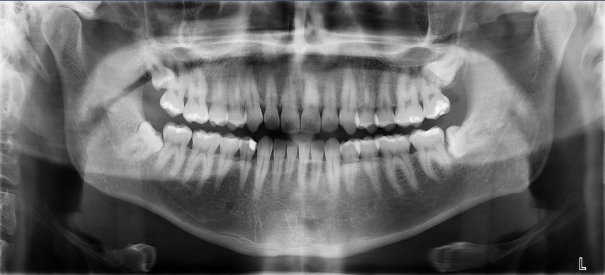
The panoramic is an overview image that is used to show the upper and lower jaw, erupted and unerupted teeth and to evaluate root alignment, TMJ’s and sinuses.

CEPHALOGRAM

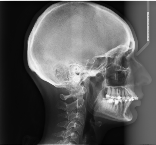
These images are acquired in standardized lateral(side) and posterior (front) projections. These views show the bone and soft tissues of the head and are used to evaluate jaw growth and spatilal relationships of the upper and lower jaws.

TMJ X-RAY

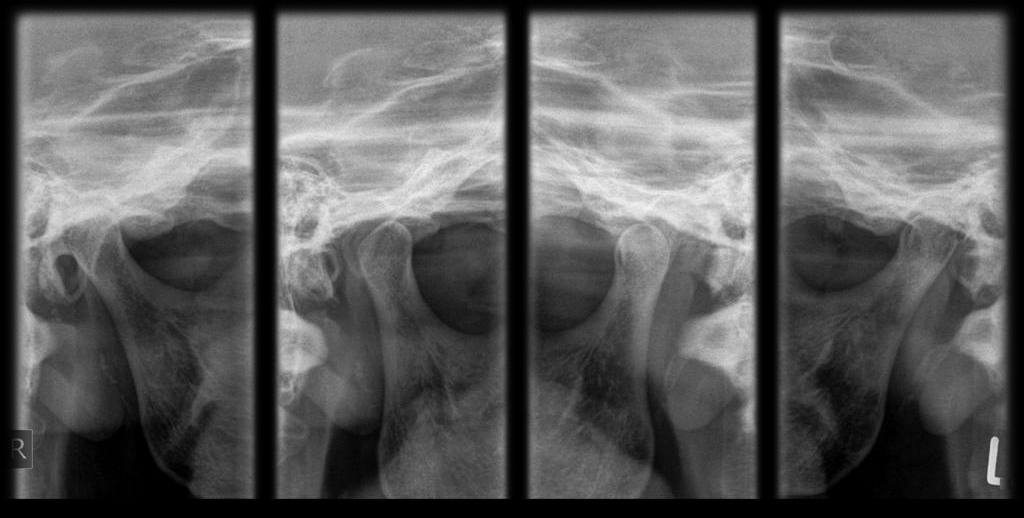
The imaging modalities is used to evaluate the anatomy of TMJ,osseus contours, condyle and range of motion.
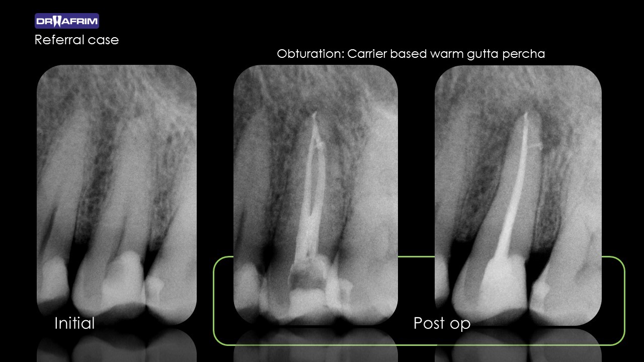
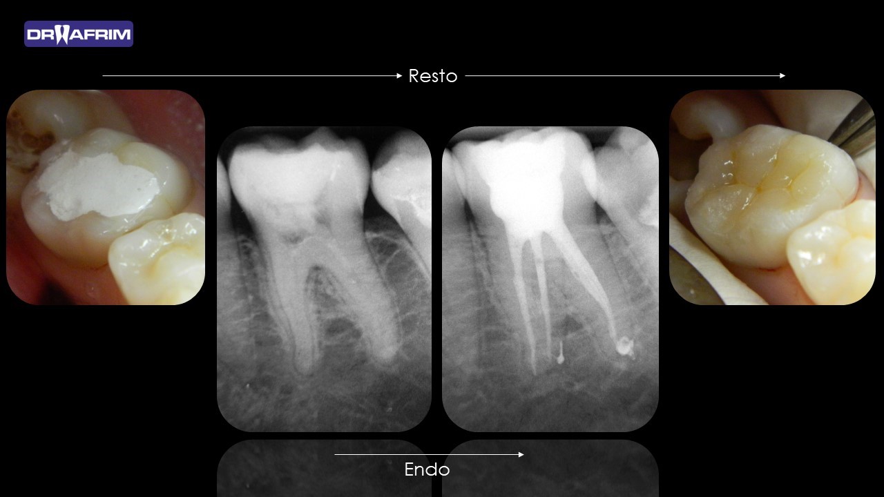
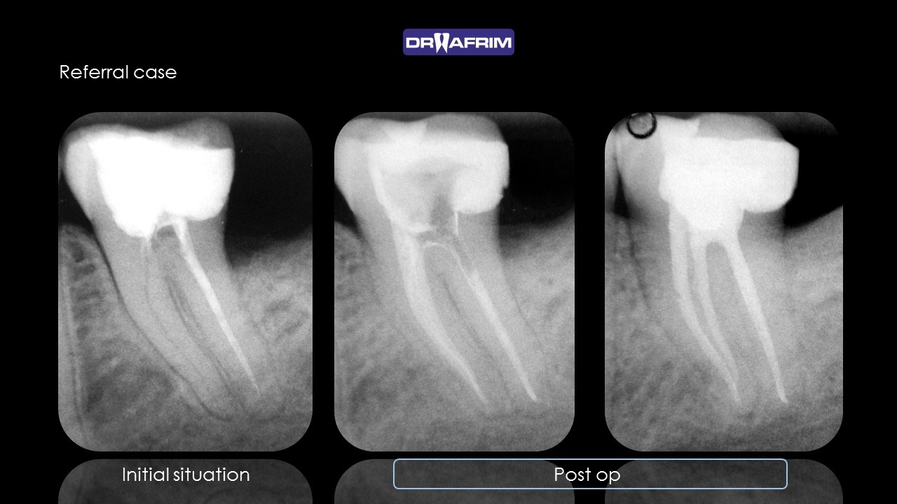
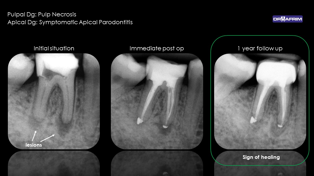
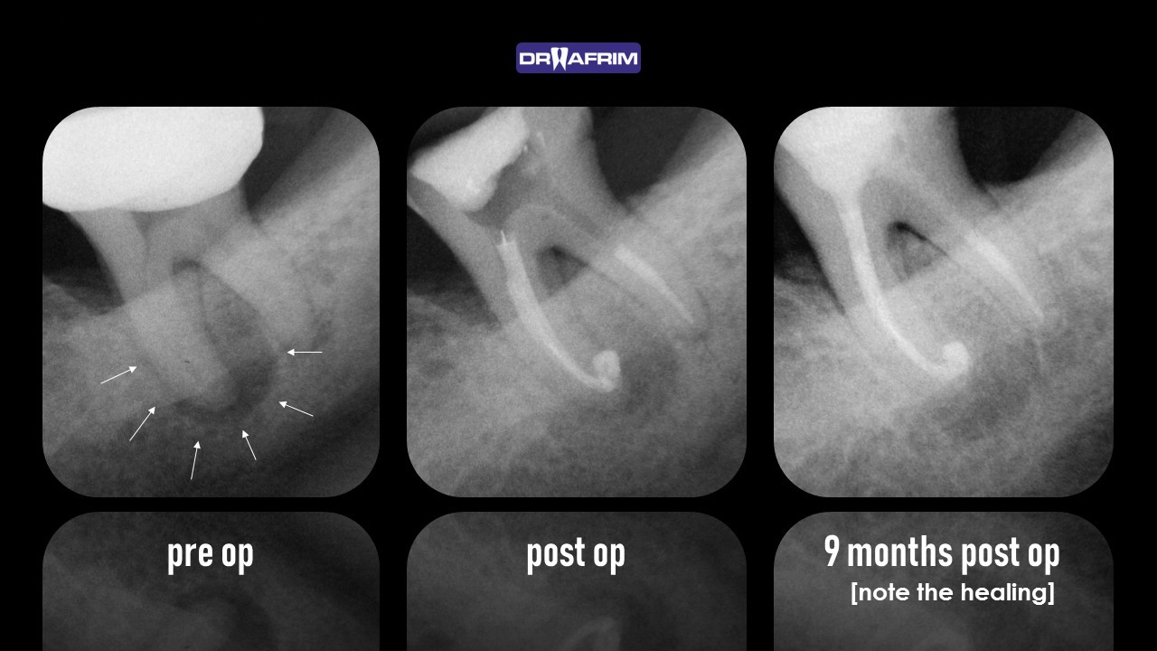
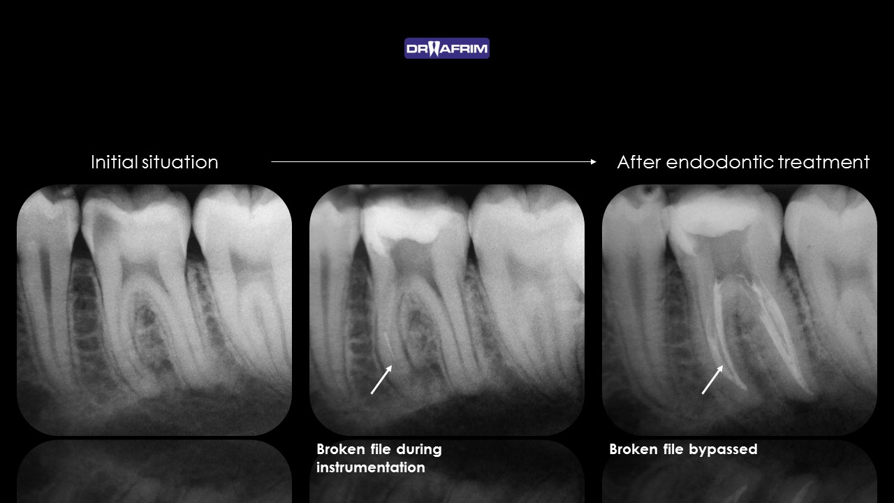
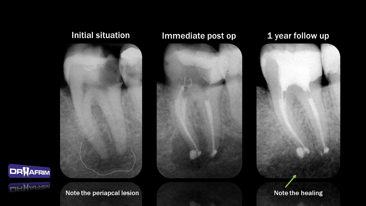
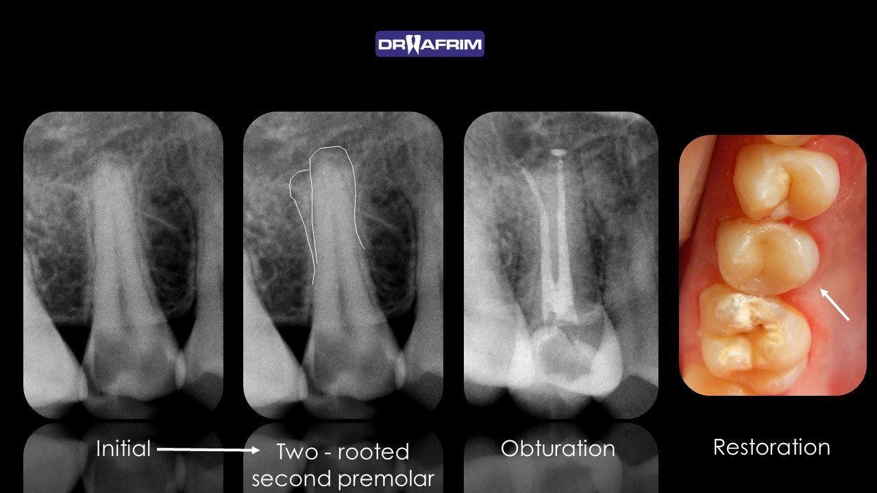
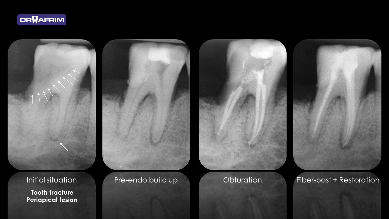
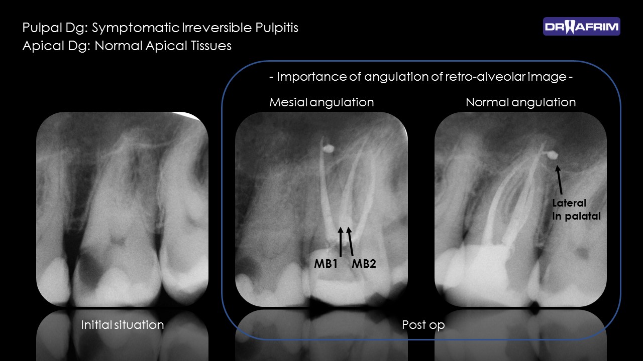
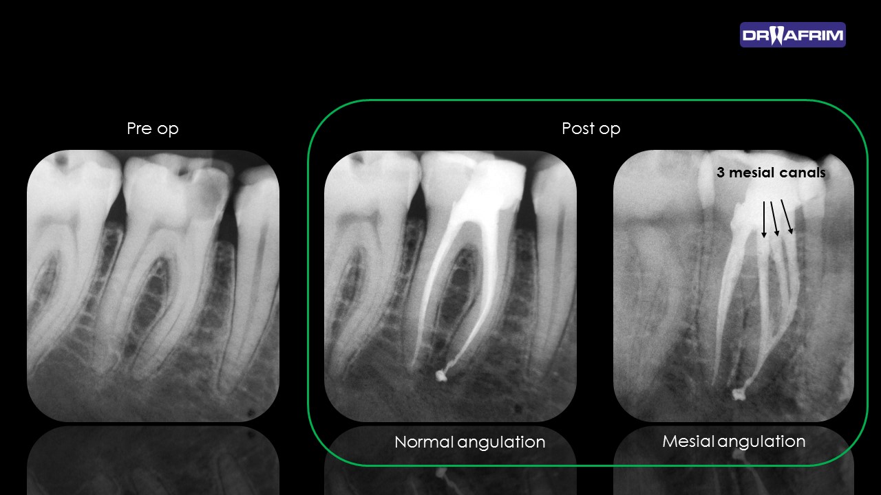
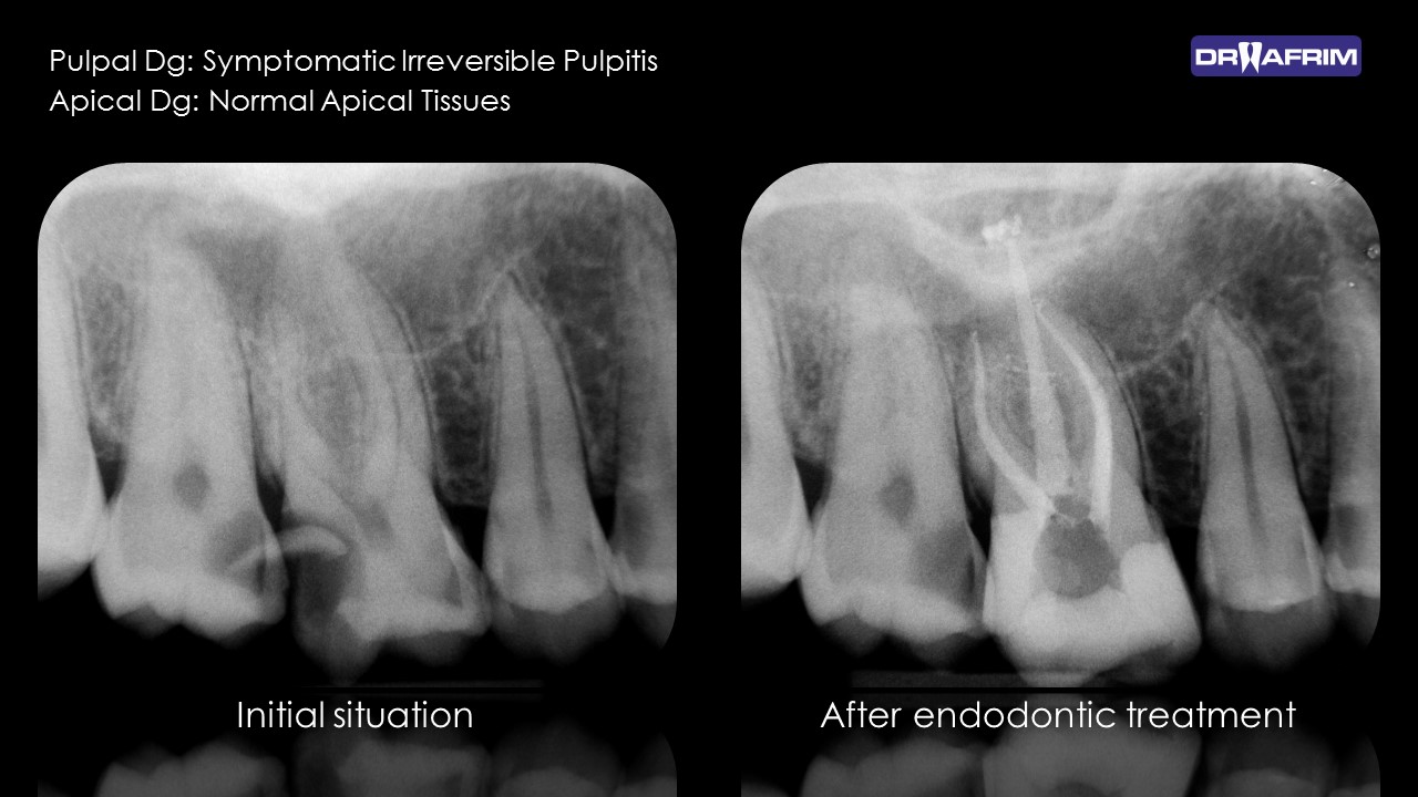
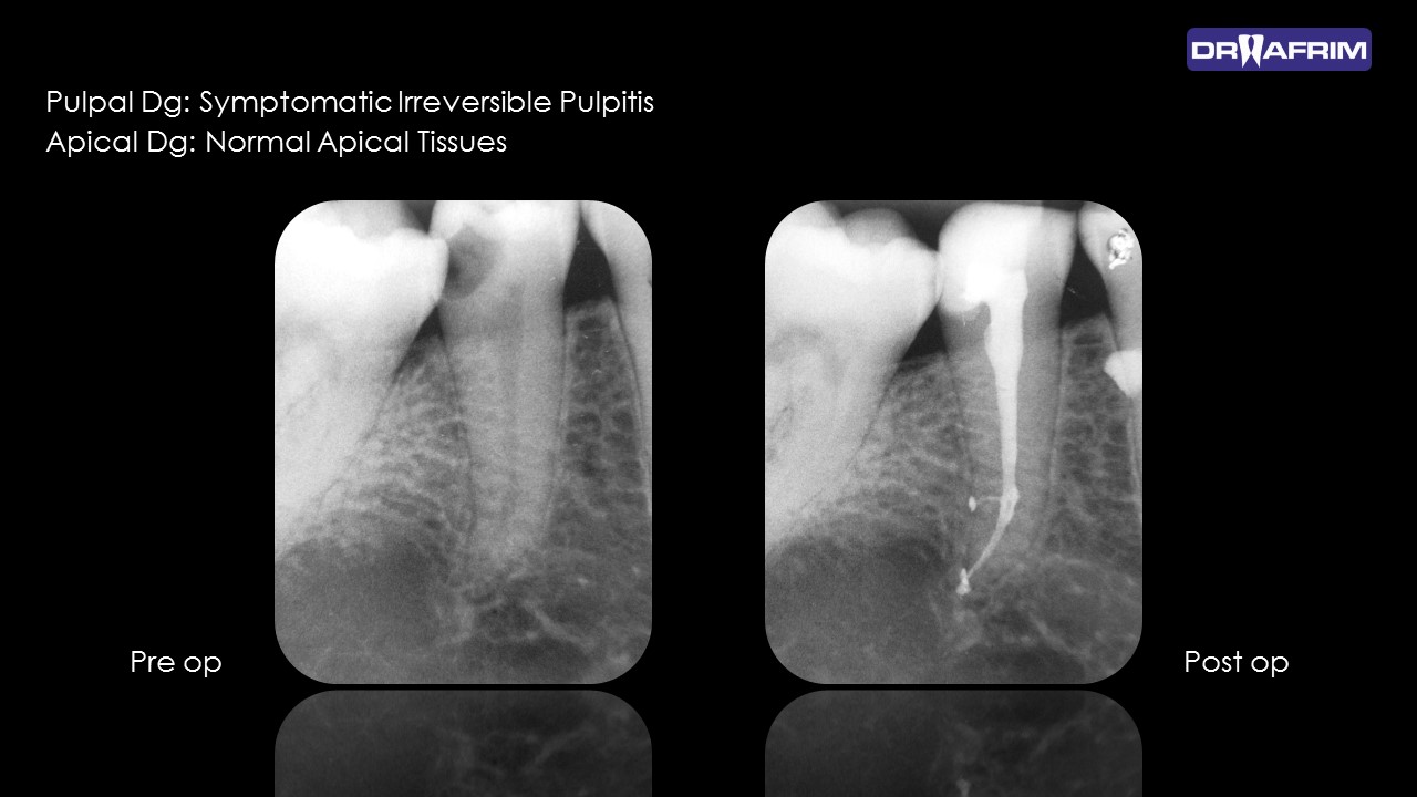
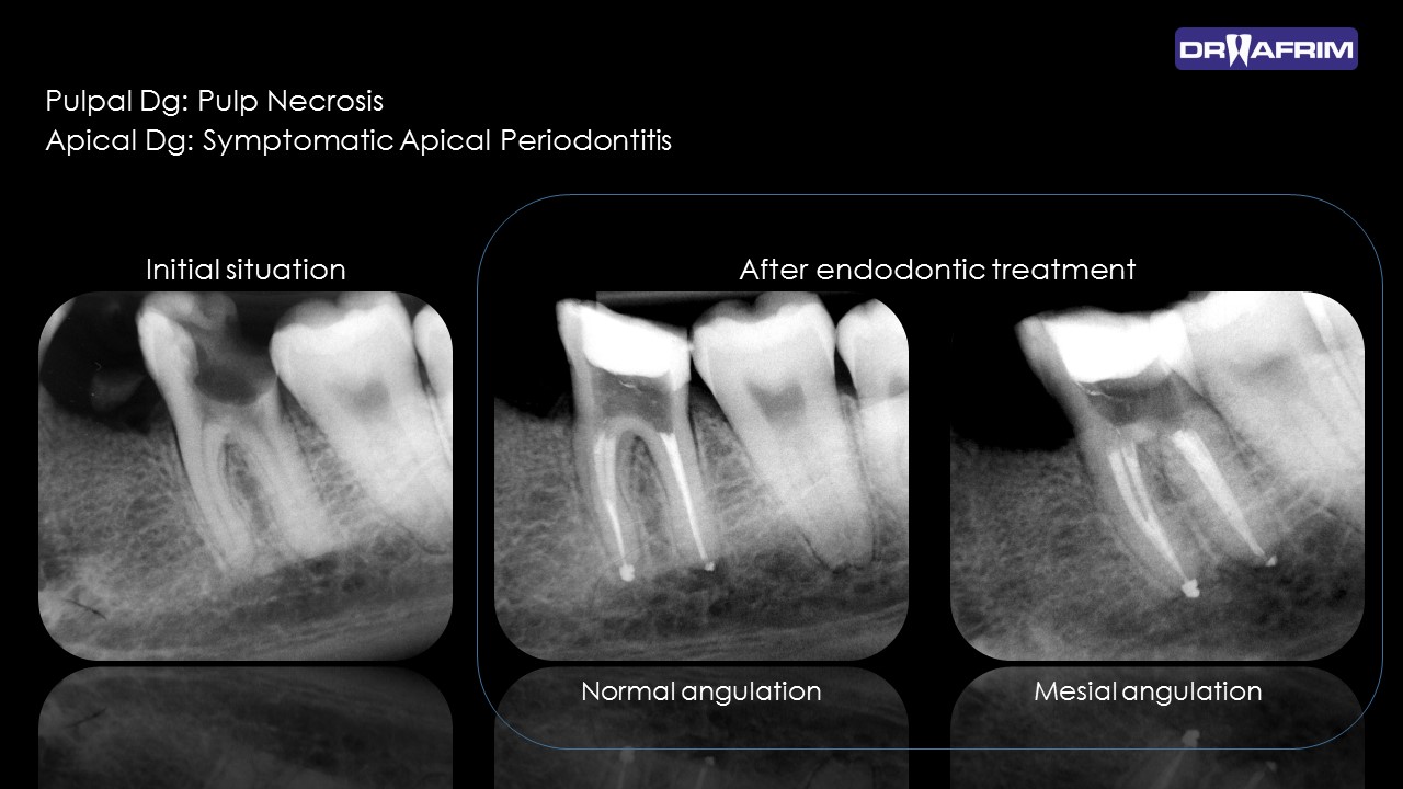
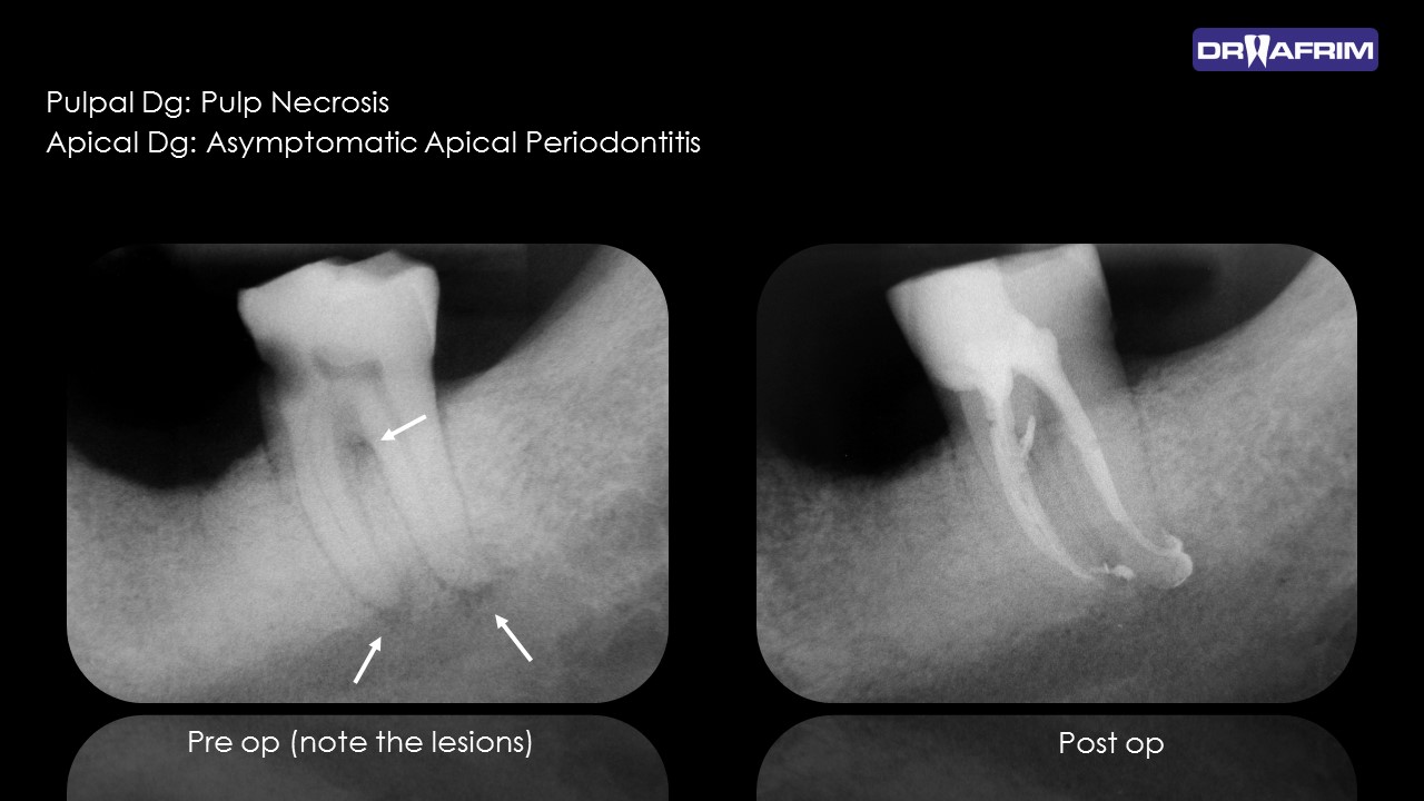
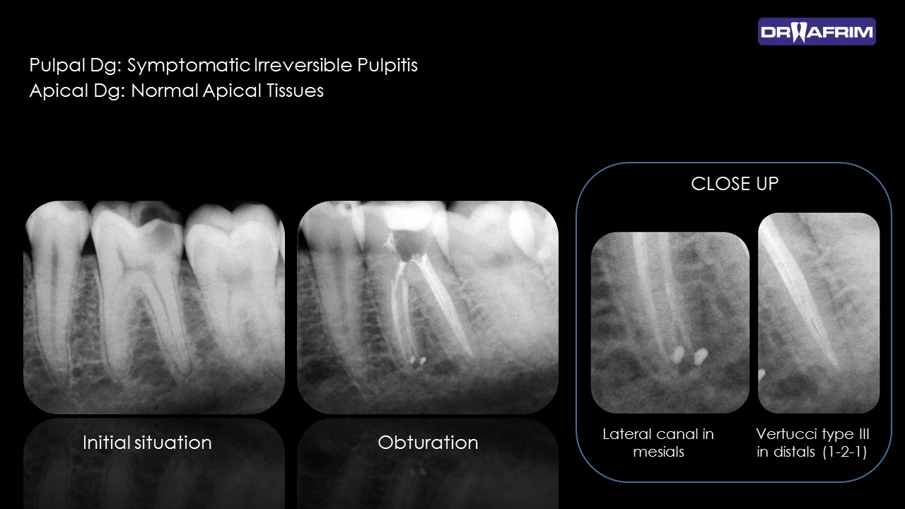
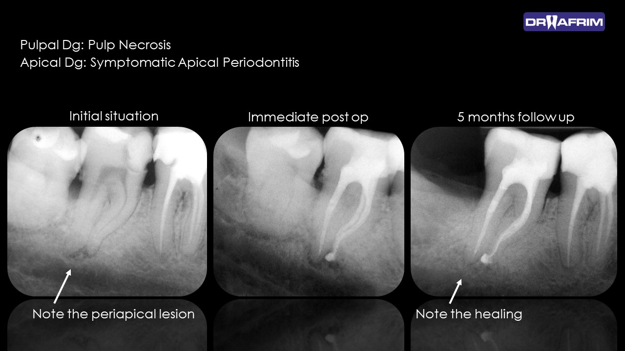
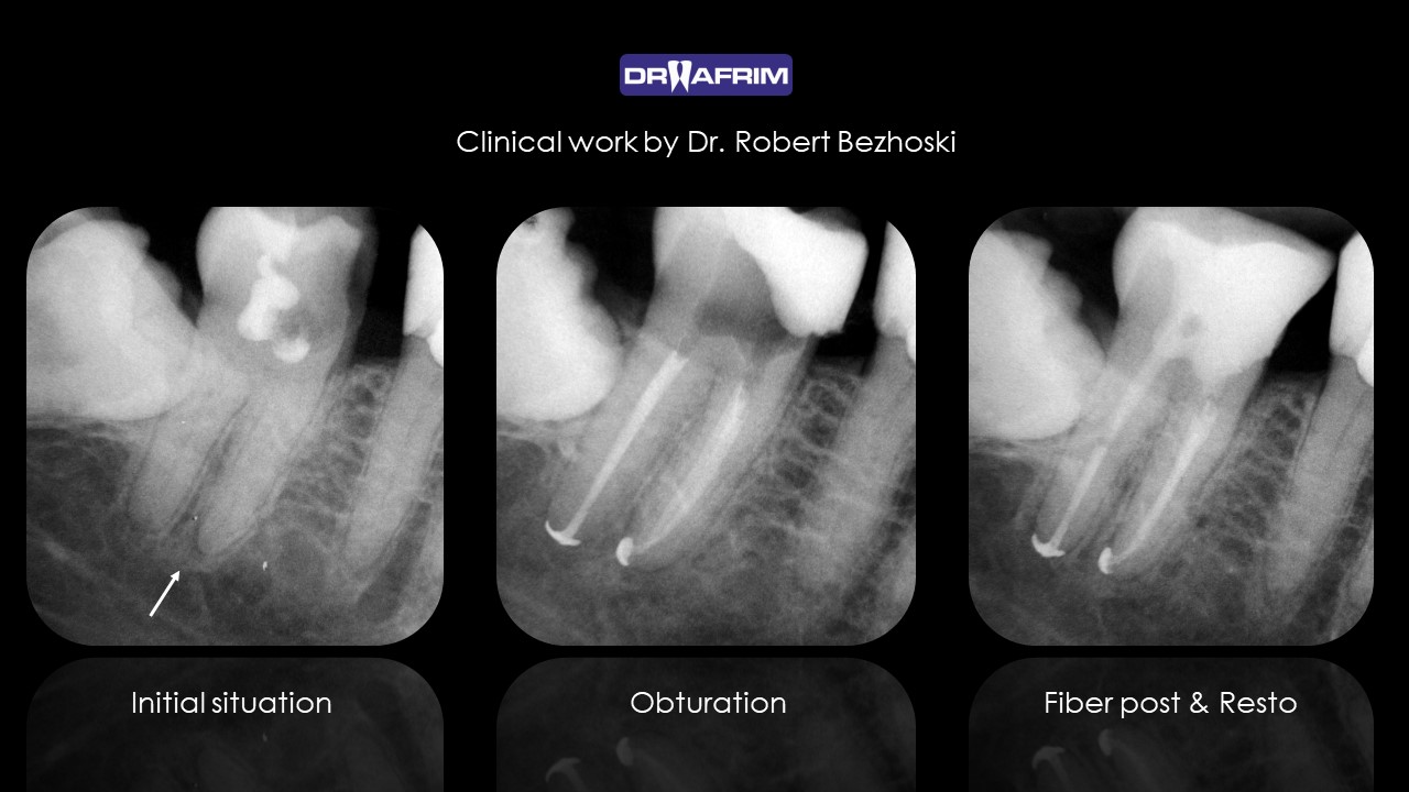
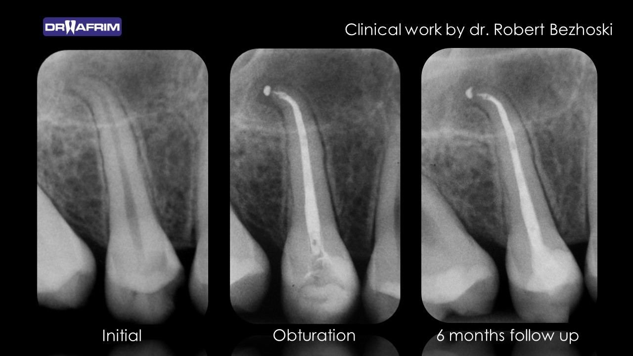
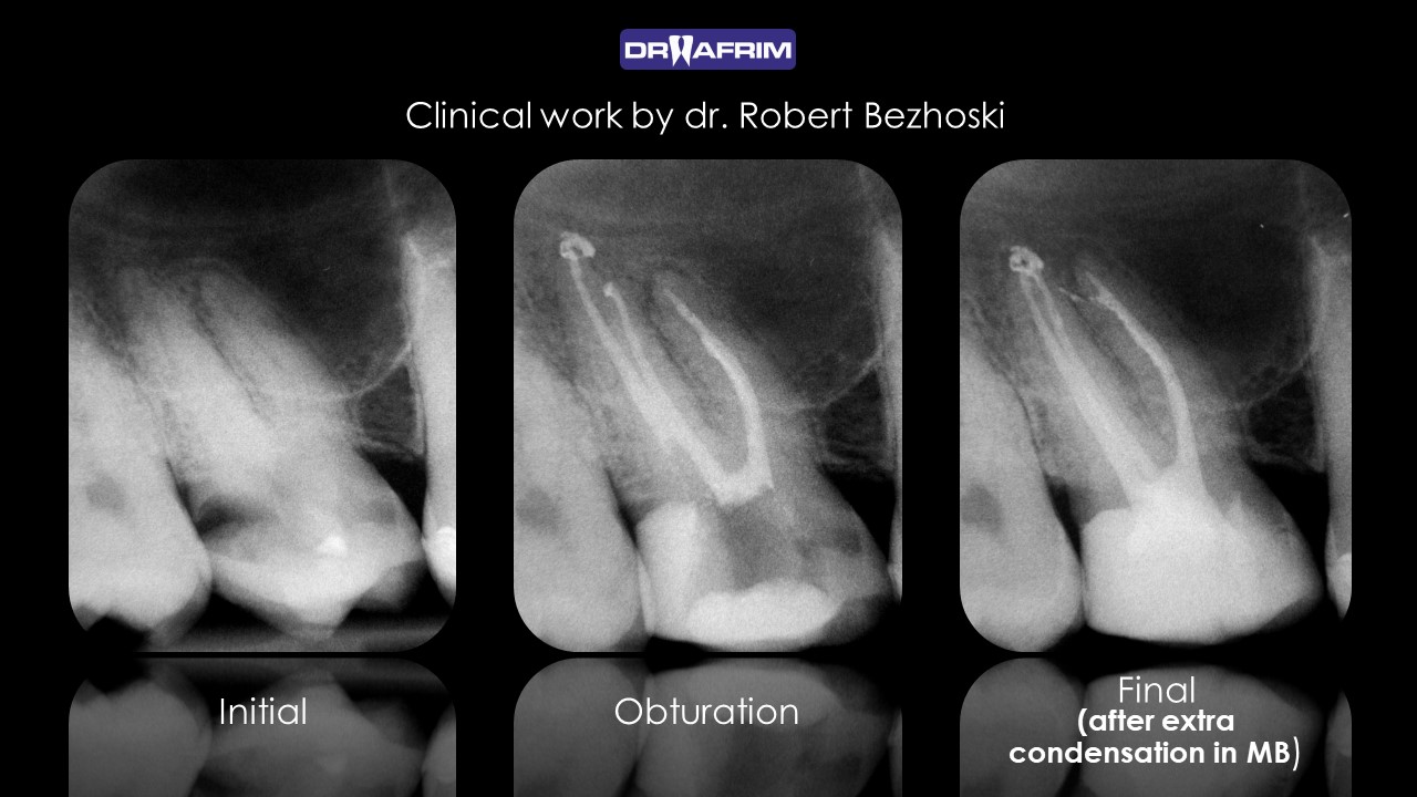
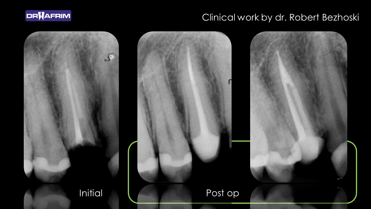
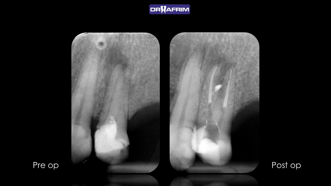
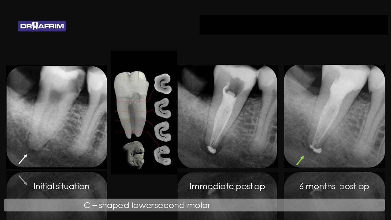
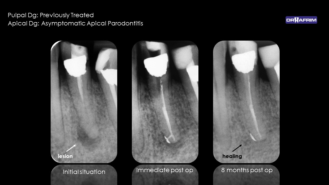
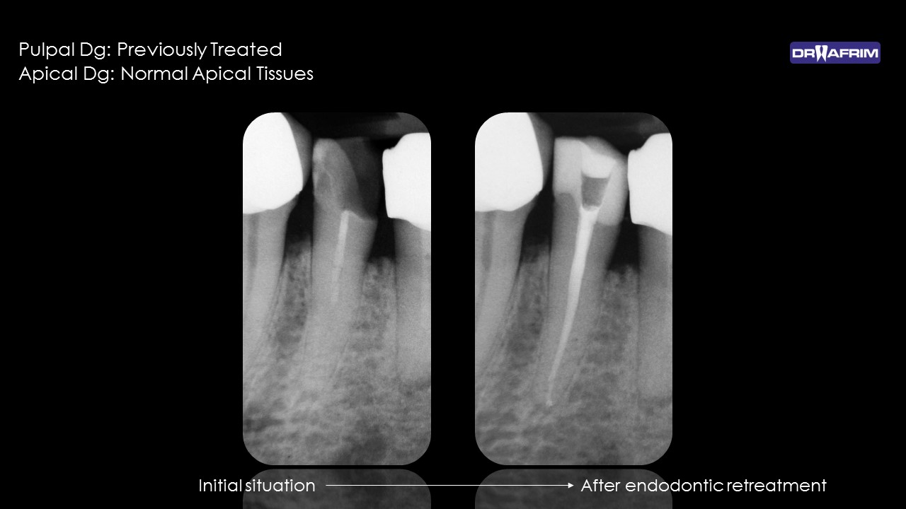
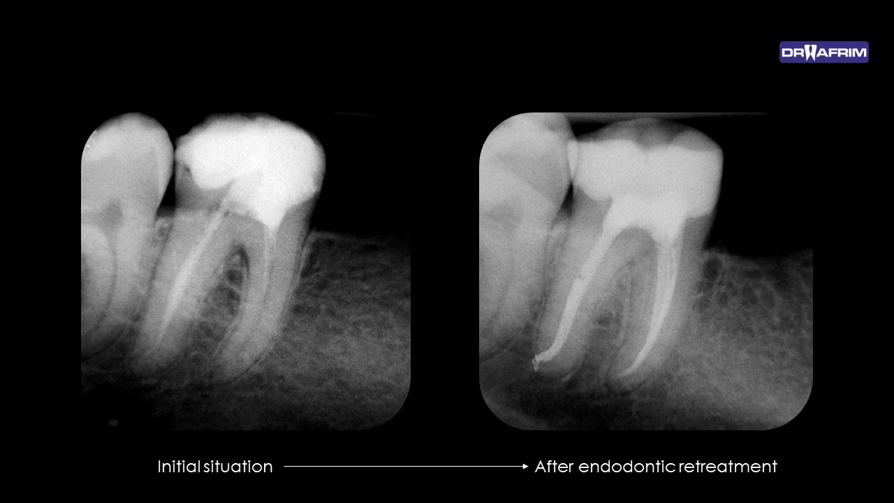
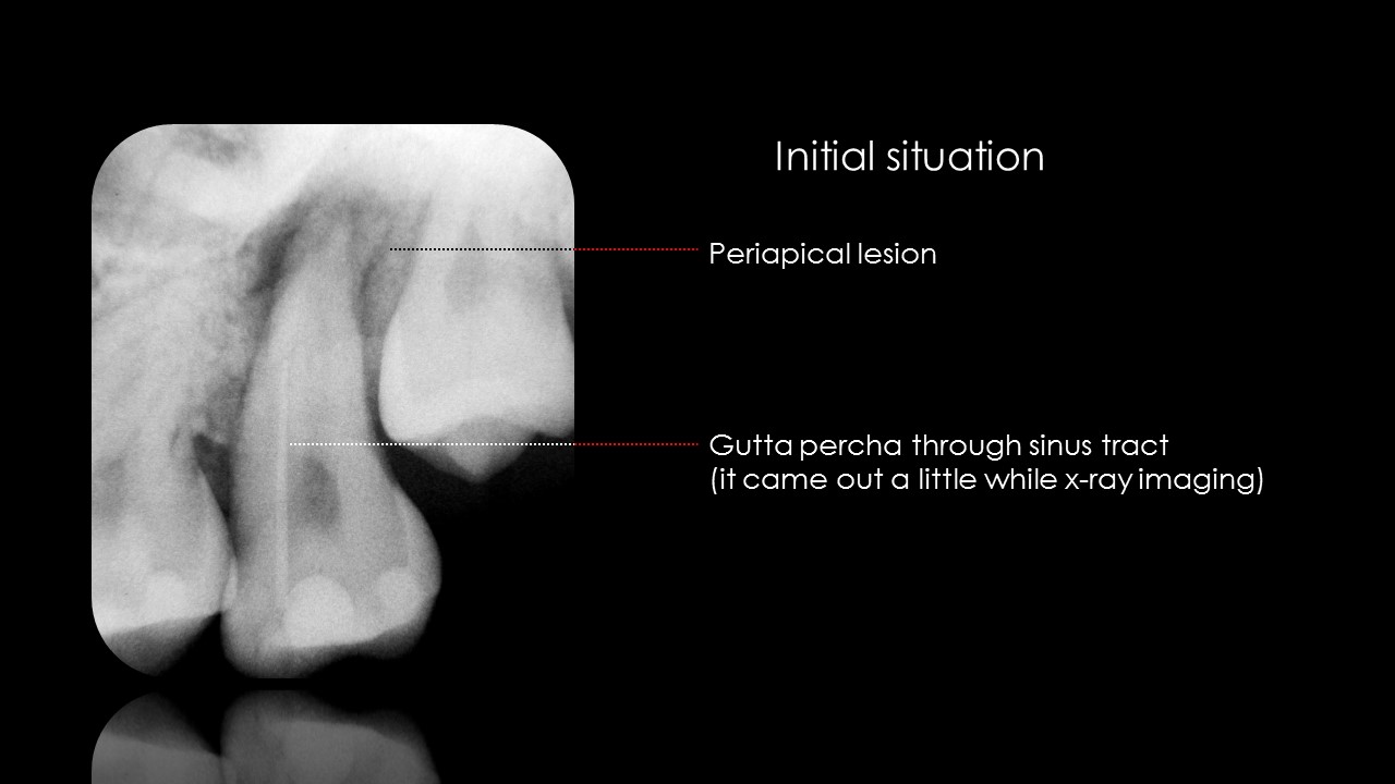
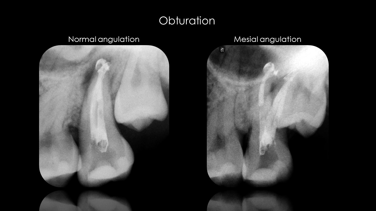
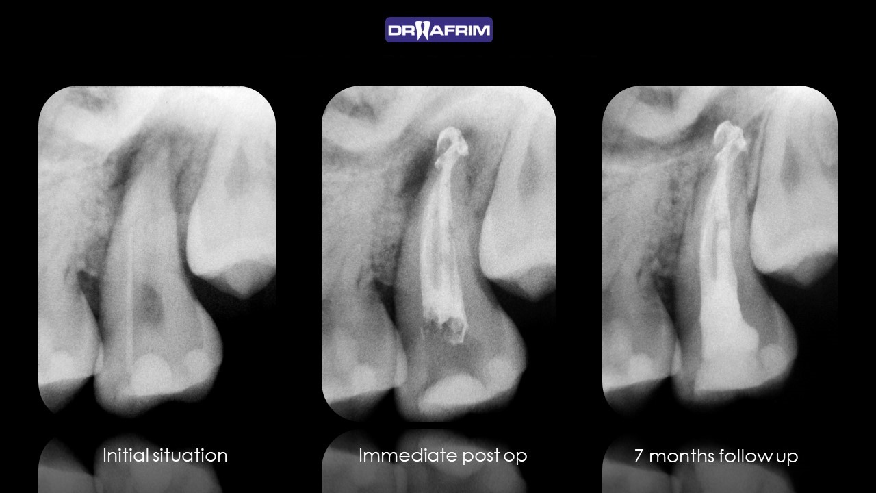
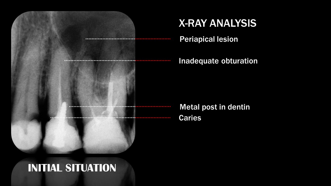
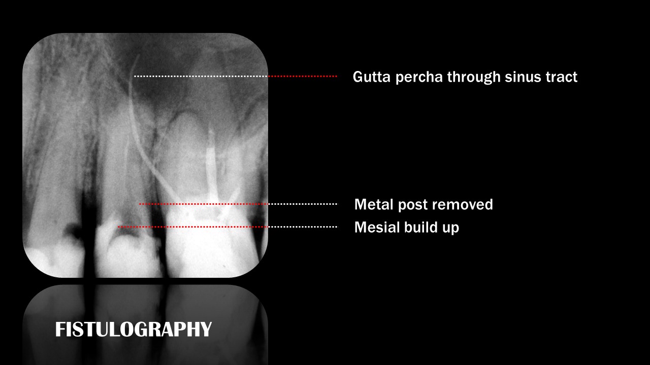
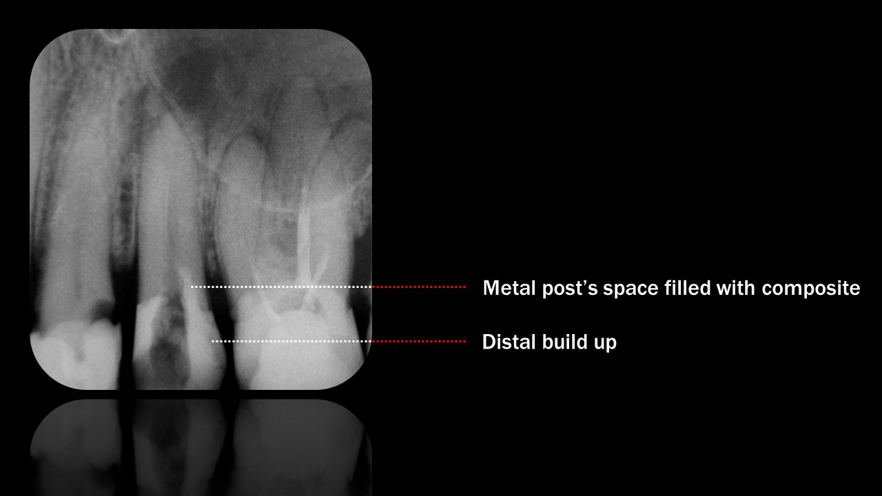
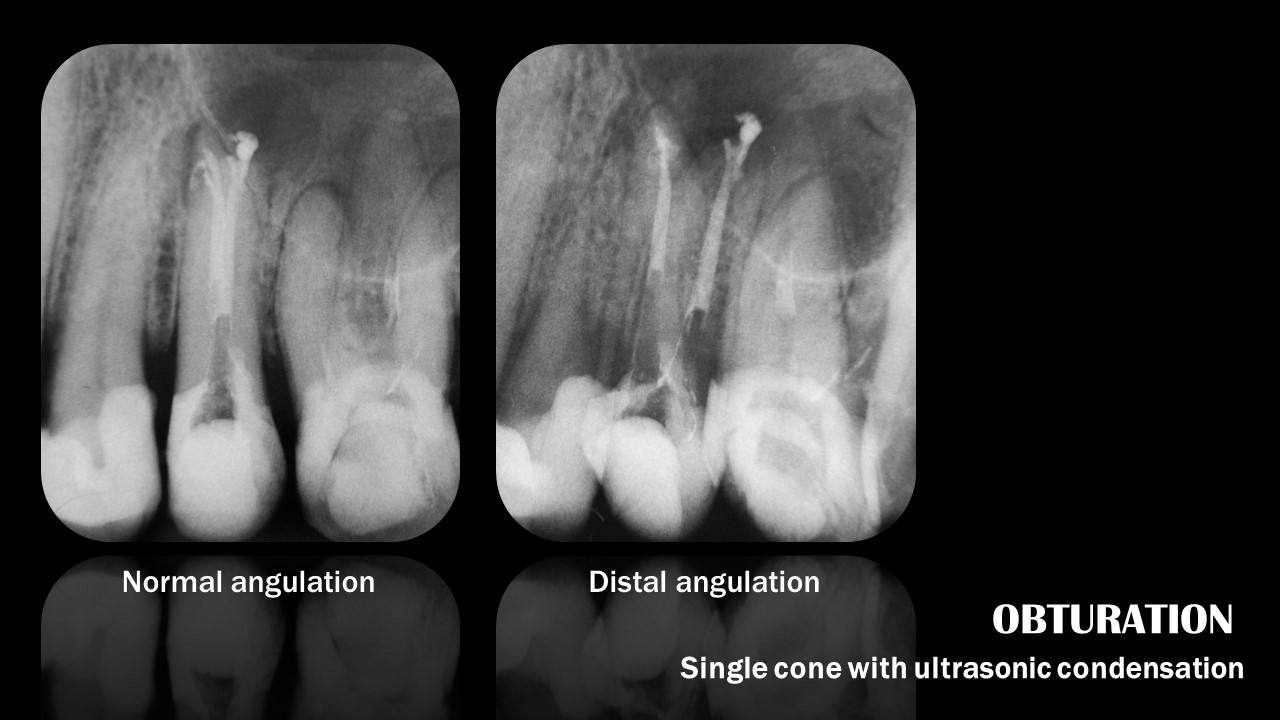
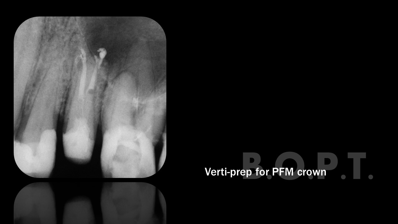
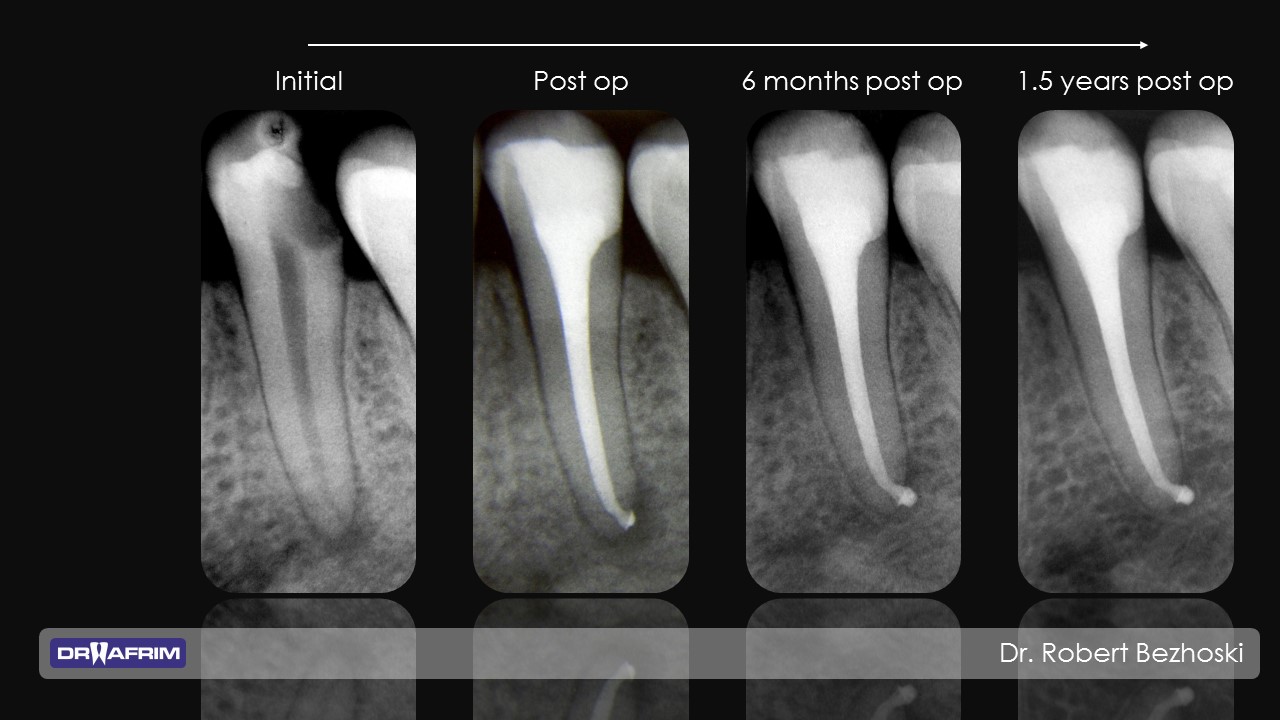
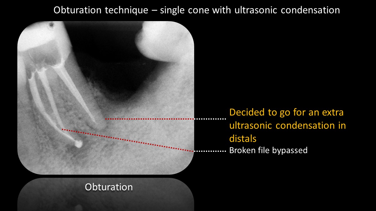
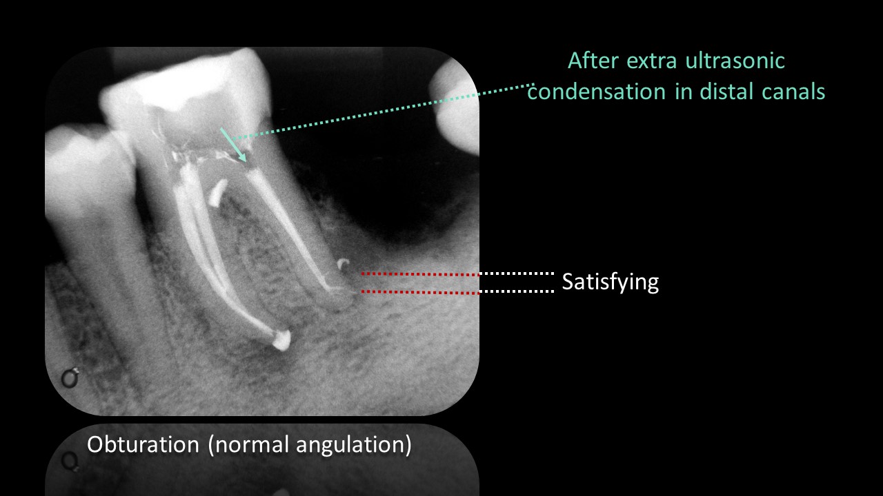
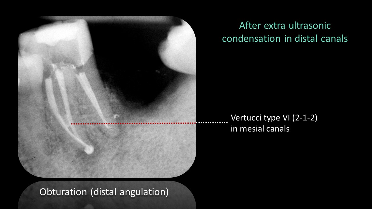
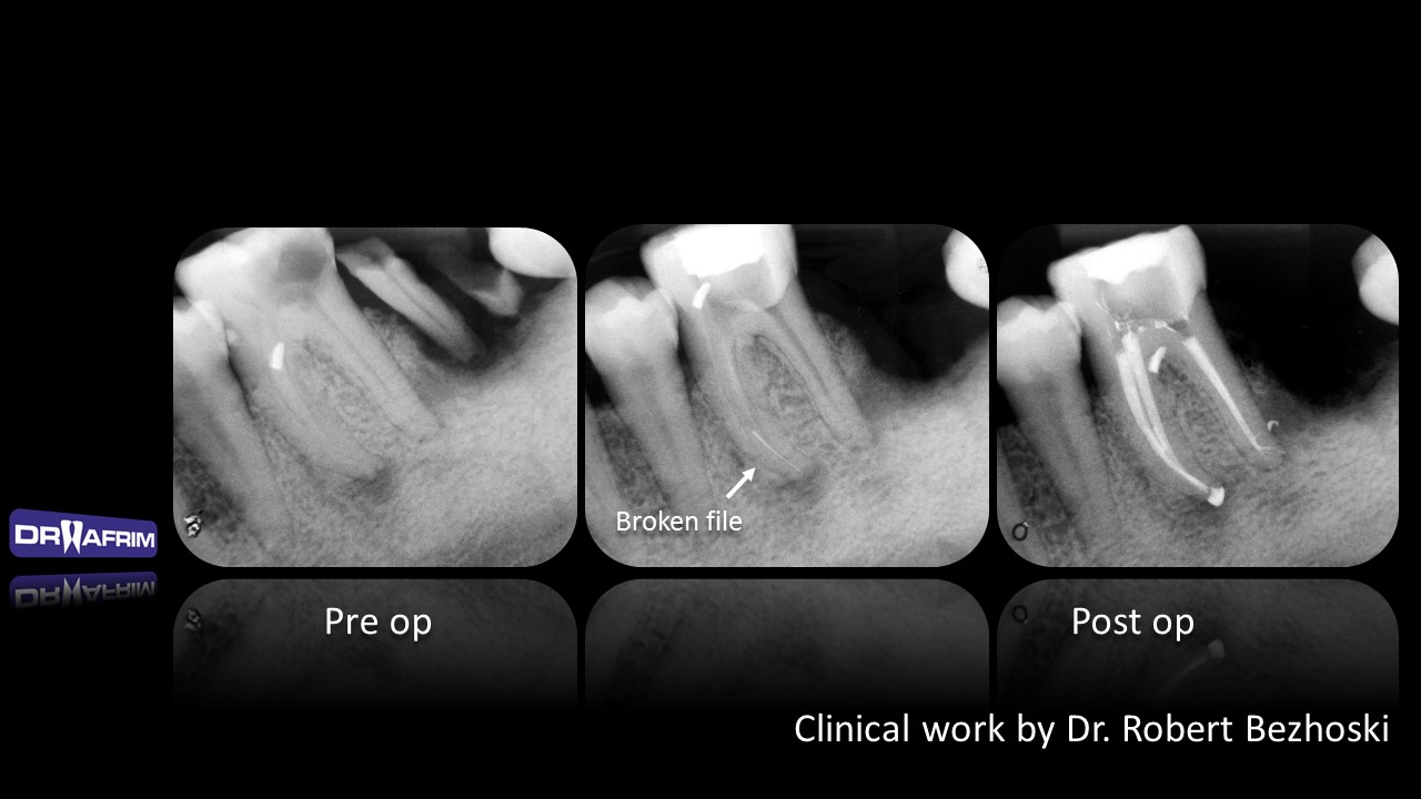
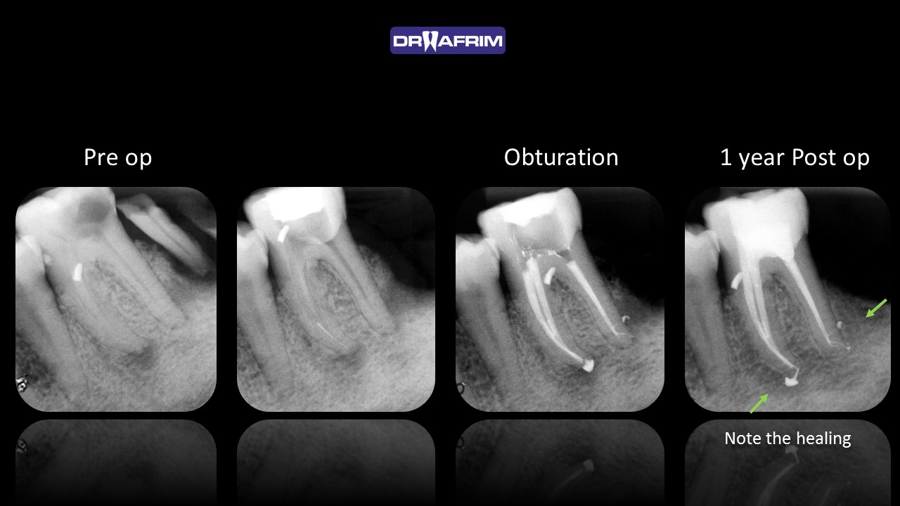
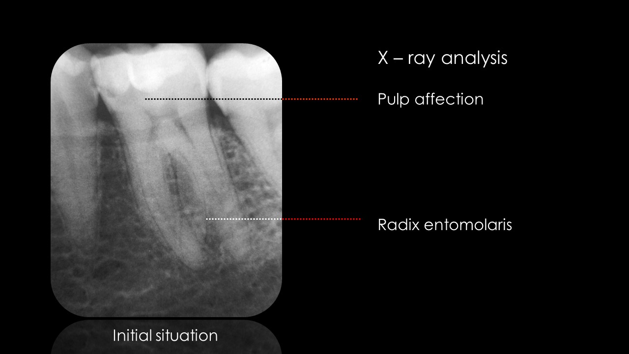
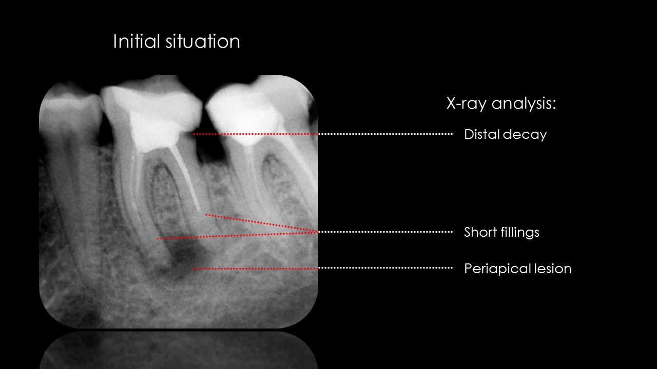
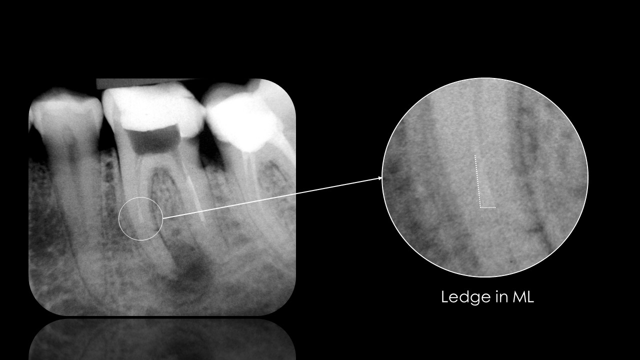
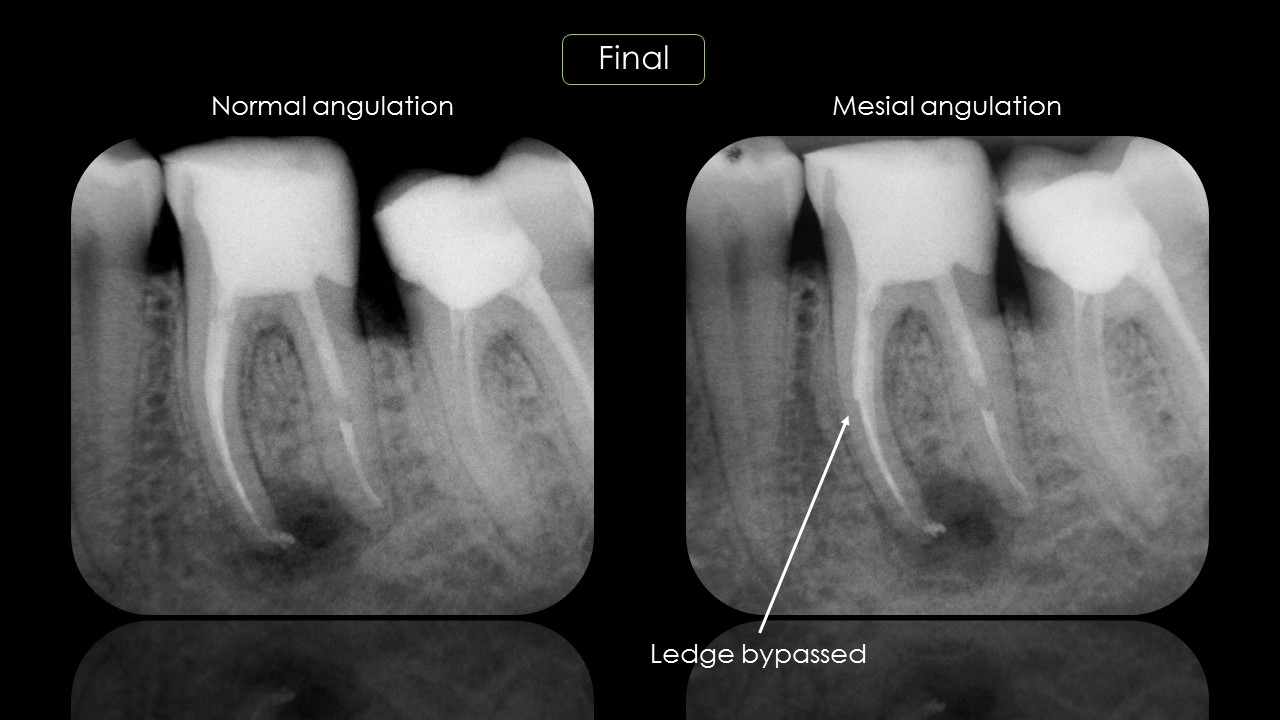
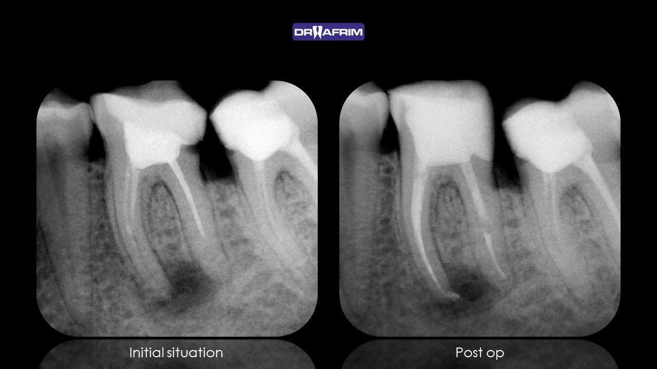
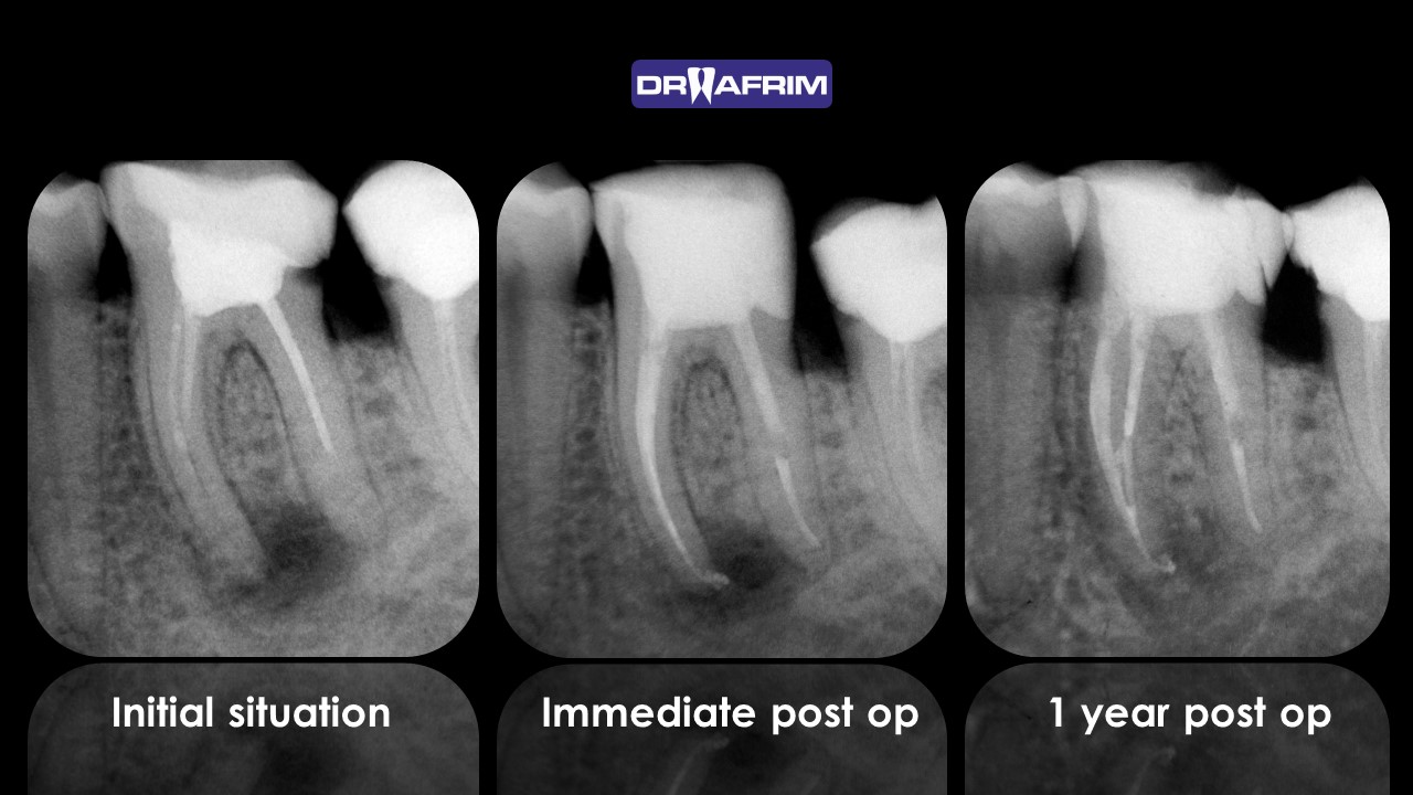
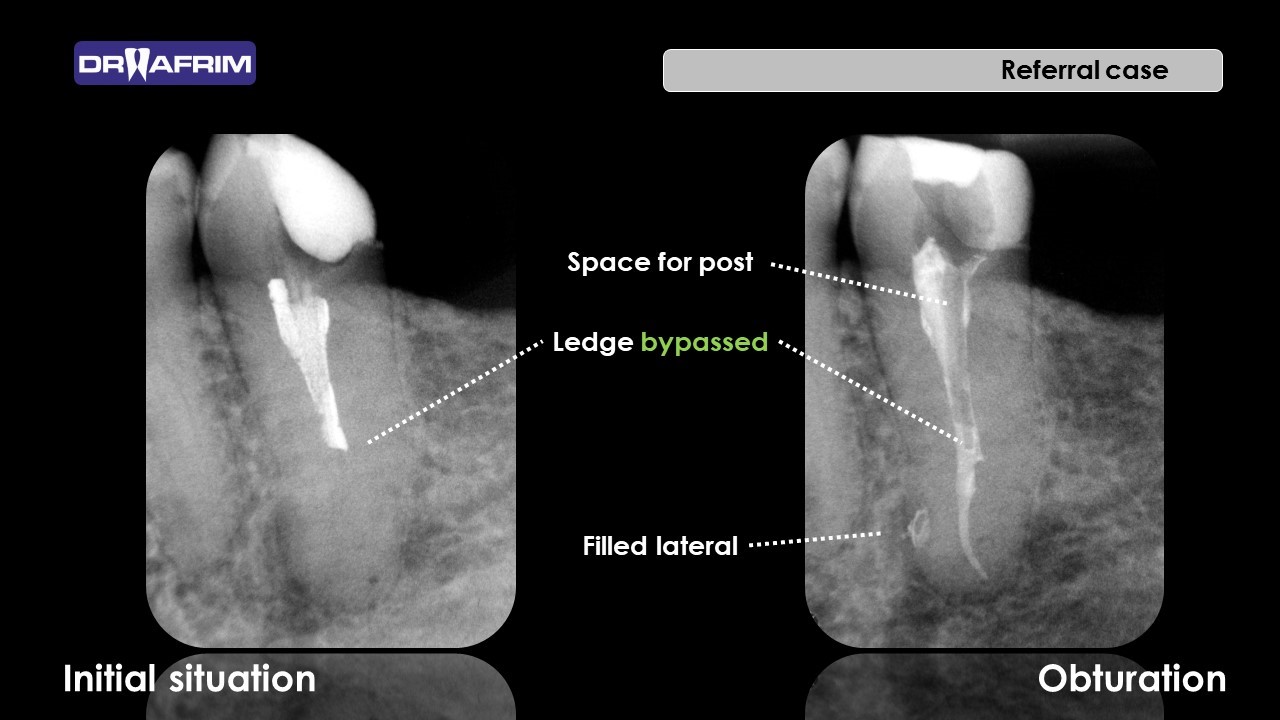
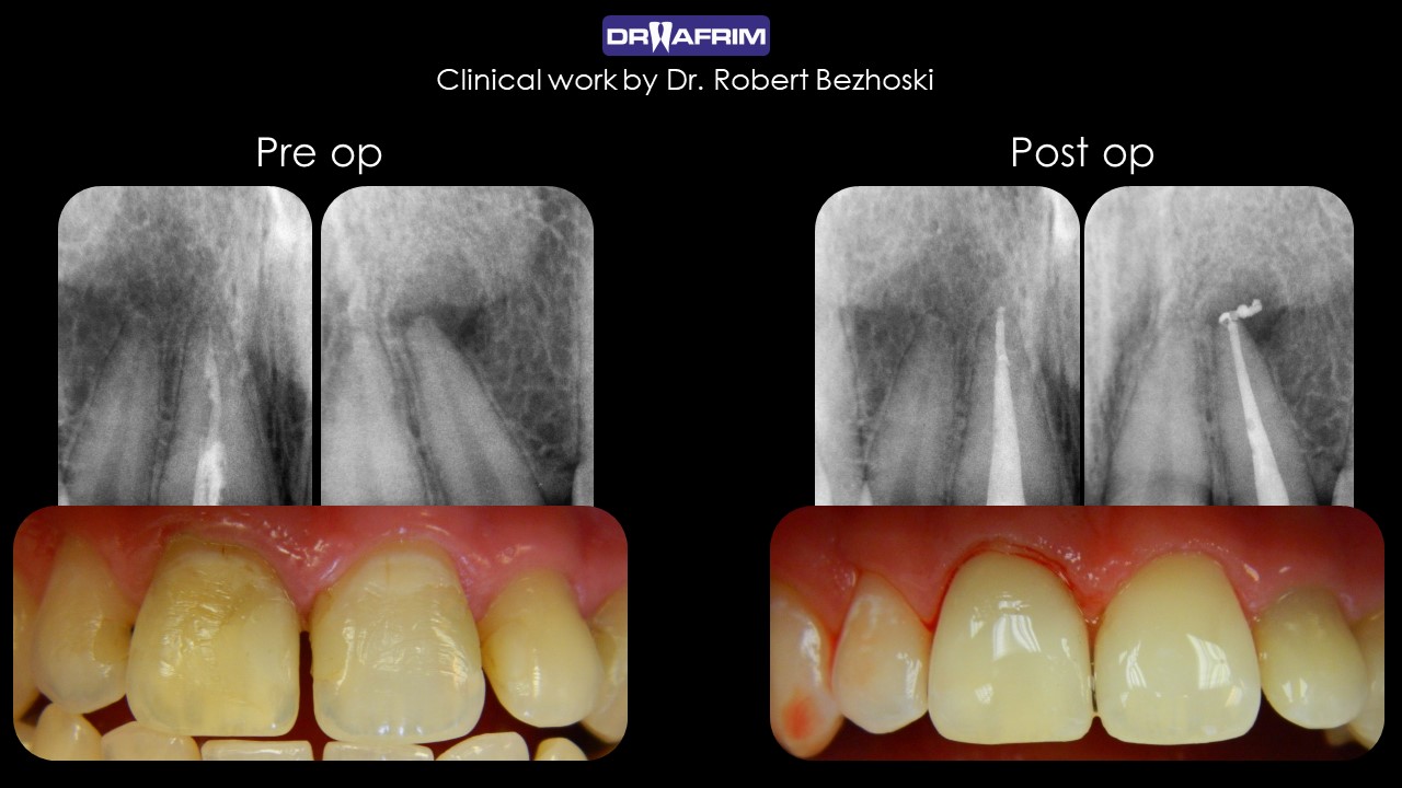
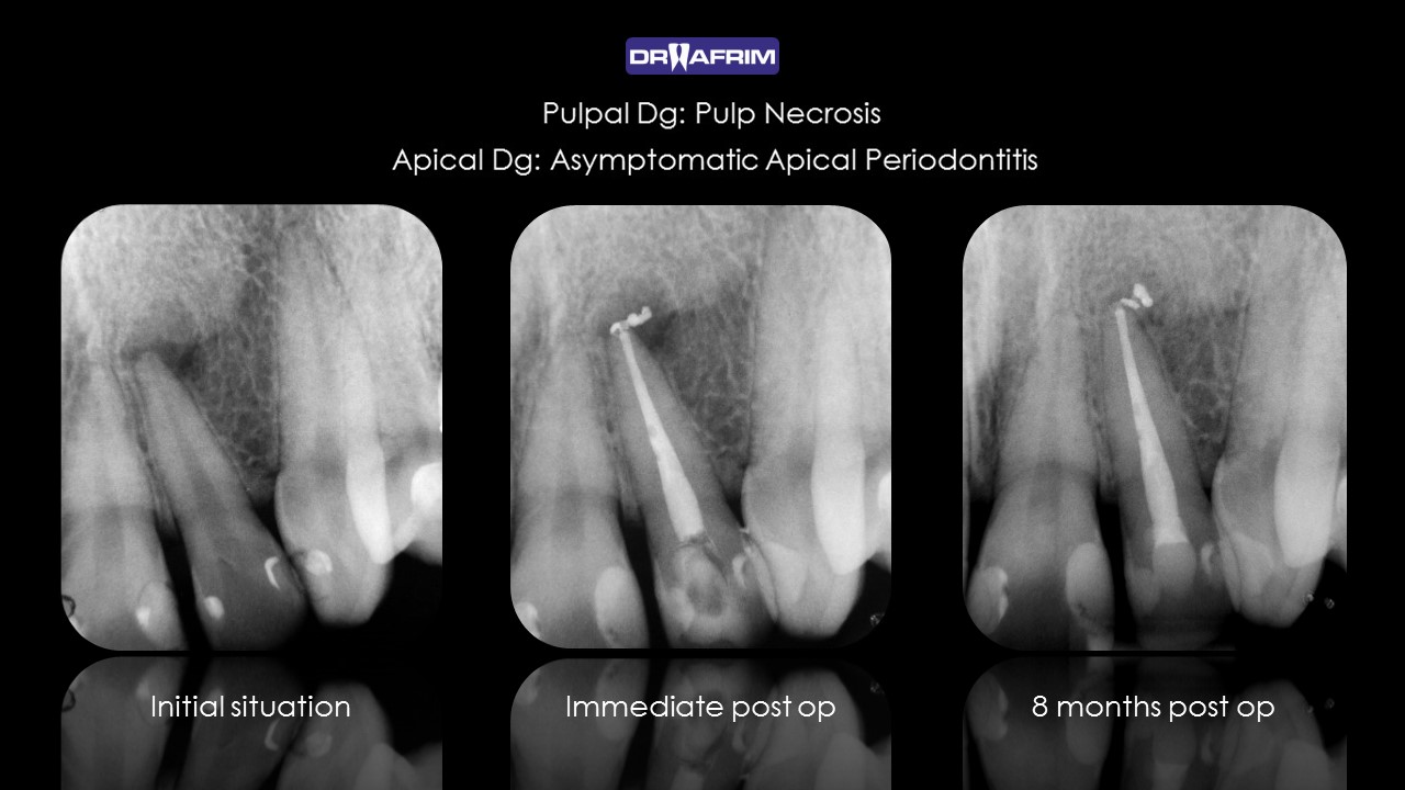
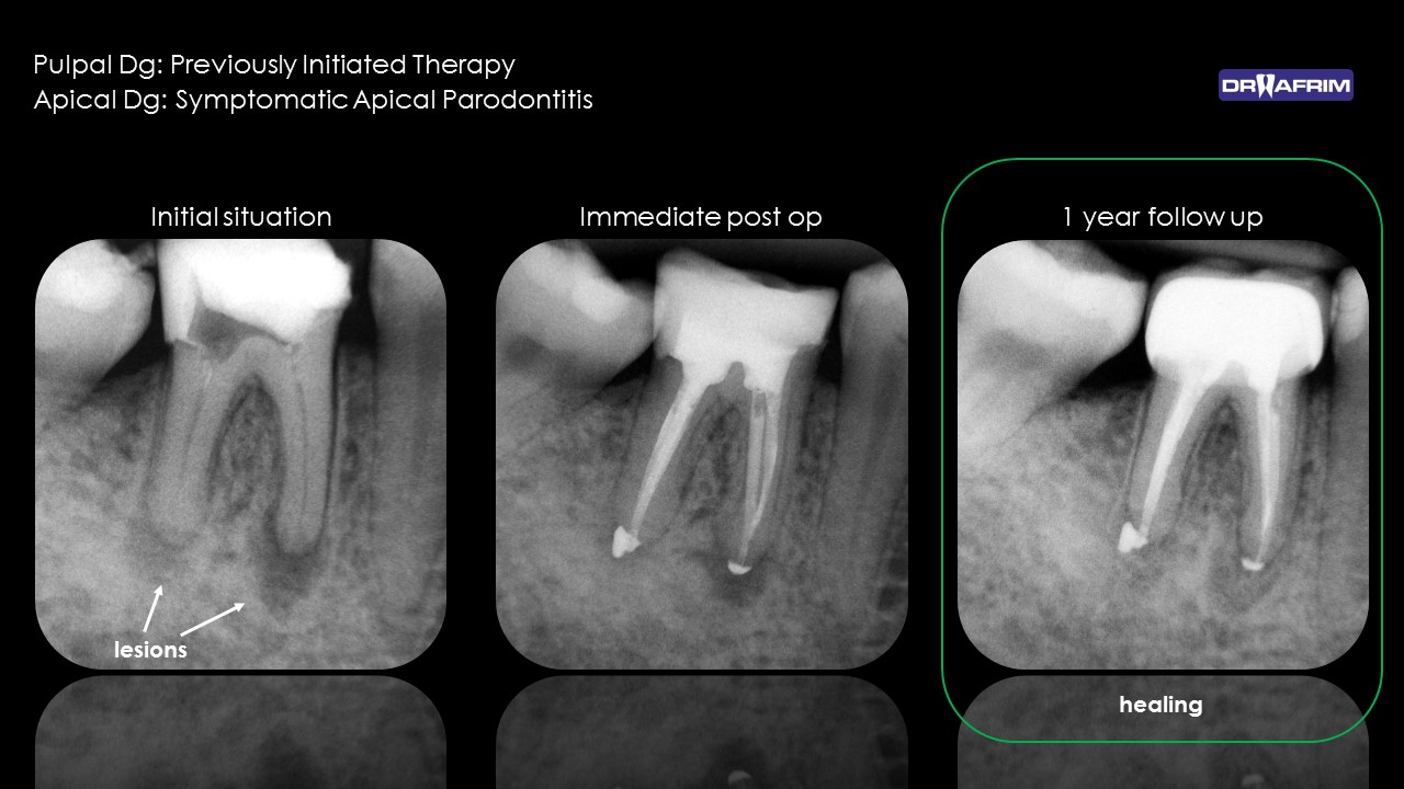
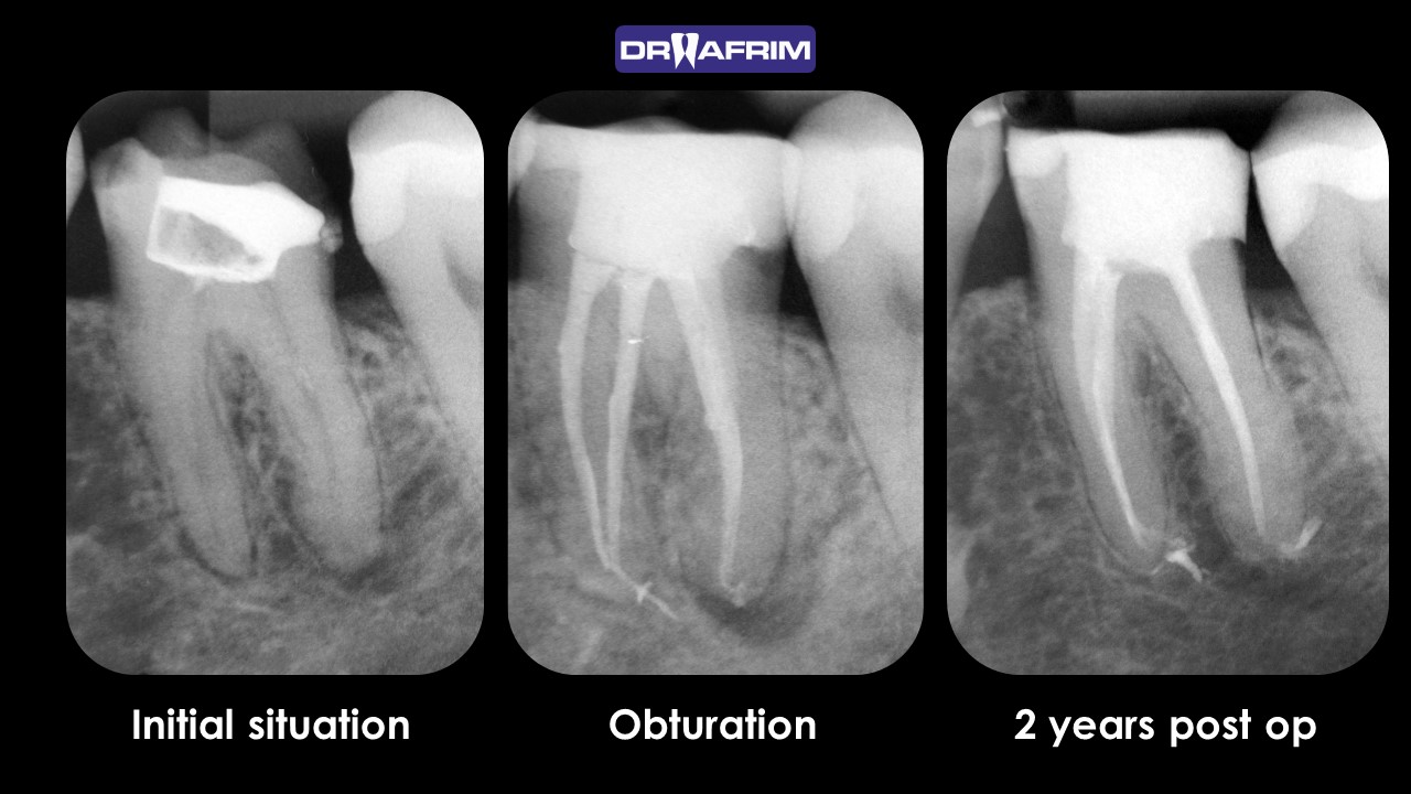
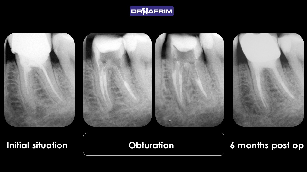
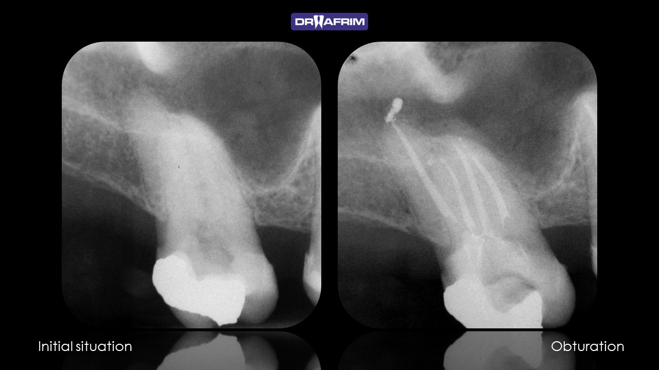
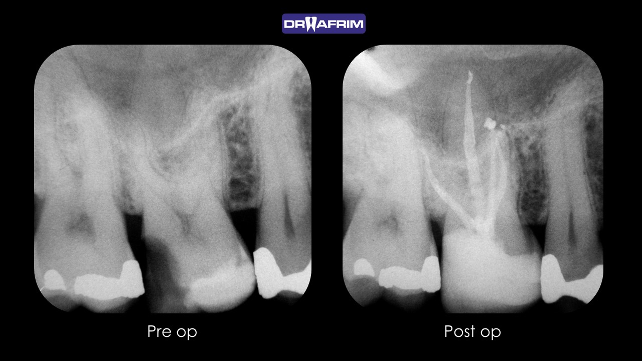
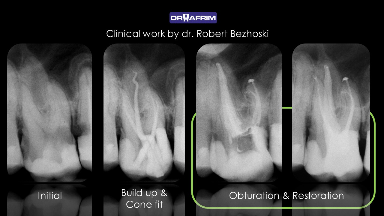
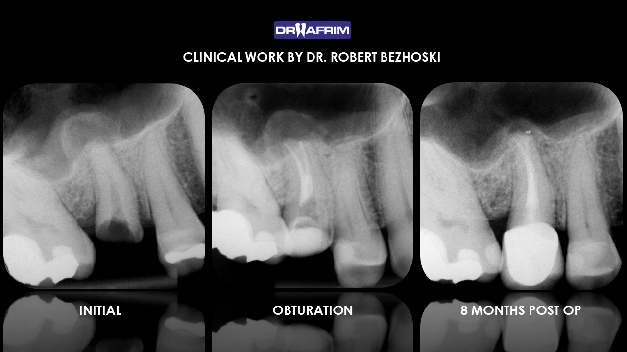
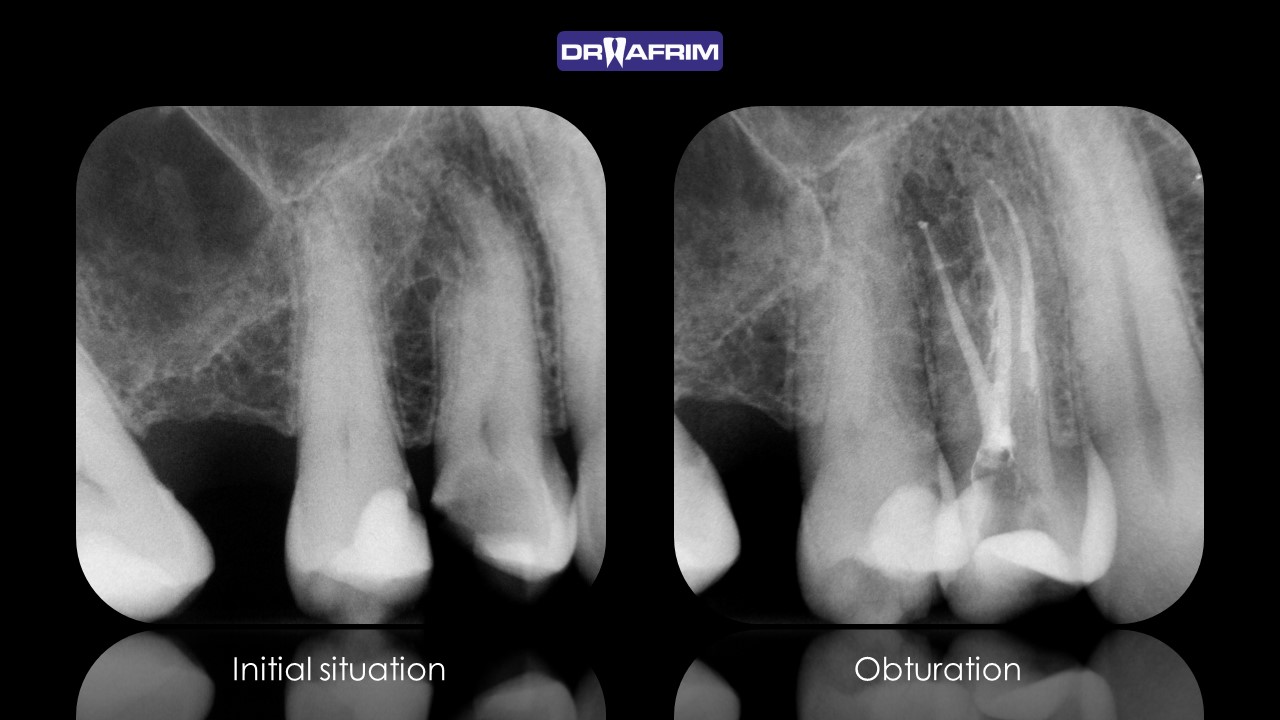
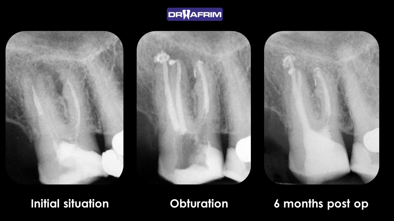
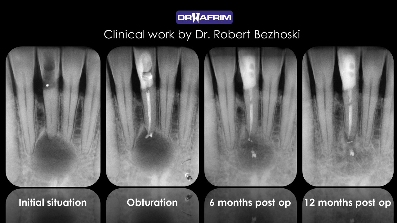
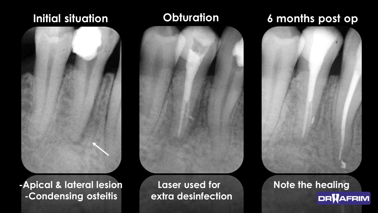
PERIAPICAL X-RAYS

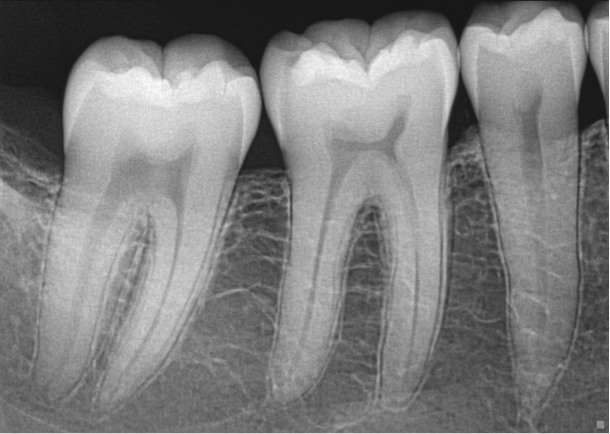
Periapical images show the teeth and supporting bone with great amount of detail. These images are used to identify dental cavities and abnormalities involving the bone adjacent to the teeth.

BITE WING X-RAYS

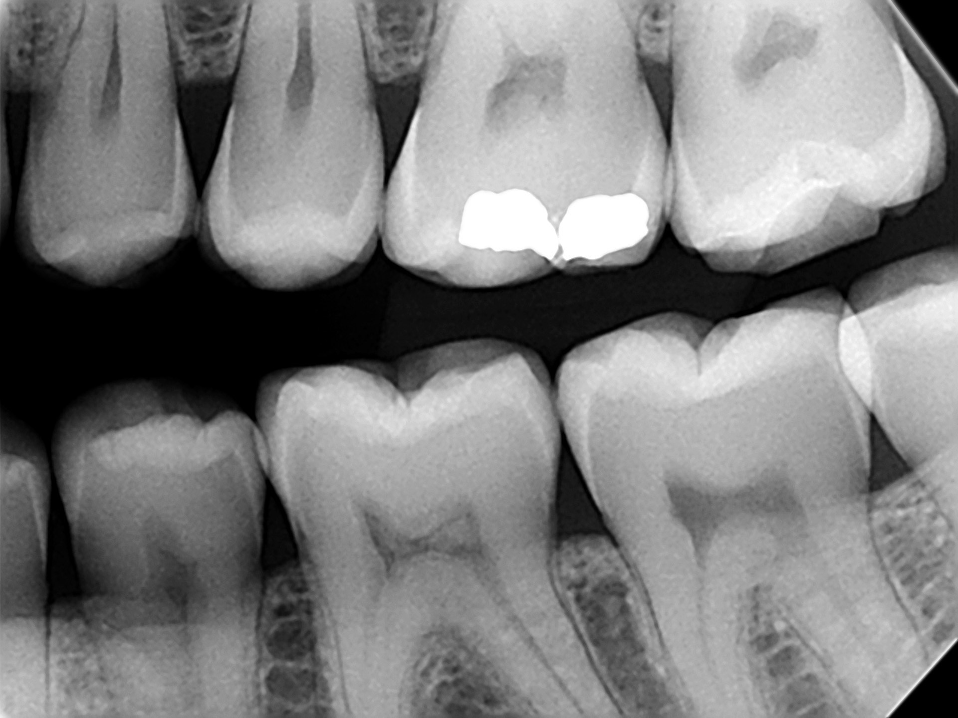
The bite wing view is taken to visualize the crowns of the posterior teeth and height of the alveolar bone in relation to the cementoenamel junctions, which are demarcation lines on the teeth which separate tooth crown from tooth root.

OCCLUSAL X-RAY

The occlusal view is indicated when is a desire to reveal the skeletal or pathological anatomy of either the floor of the mouth or the palate.

FULL MOUTH SERIES

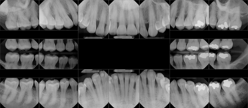
Full mouth series is a complete set of intraoral x-rays taken of a patient’s teeth and adjacent hard tissue. The full mouth series is composed of 18 films.

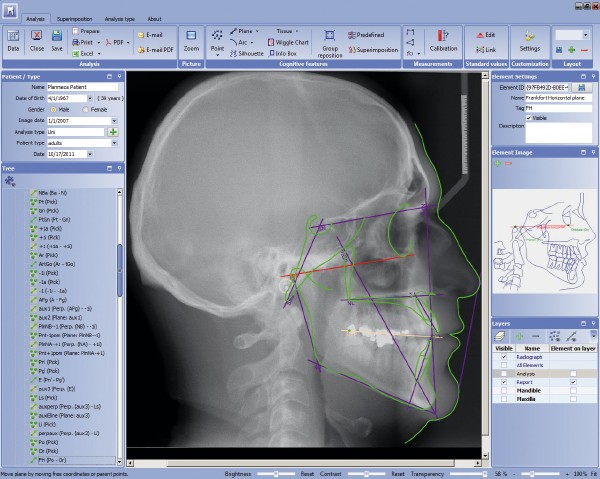
CEPHALOMETRIC ANALYSIS

3D

40 x 50

100 x 60

100 x 100

200 x 170

Clinical cases

Endodontics is branch in dentistry that involves treatment of the root canals of teeth that have been damaged by severe decay, infection or trauma.

Clinical case of the month

FEBRUARY

MARCH

Teeth with large periapical lesions does not necessary need to be submit to surgery or extraction. Endodontic treatment is conservative approach which can lead to successful healing, without surgical trauma or losing the tooth. Regular follow up is necessary.

APRIL

MAY

JUNE

CBCT before endodontic therapy enables good planning and analysis with predictable outcomes. Especially helps in multi-rooted teeth, preventing from over-instrumentation, creating of ledge or separation of files.

JULY

AUGUST

SEPTEMBER

OCTOBER

NOVEMBER

DECEMBER

JANUARY 2020

FEBRUARY 2020

MARCH 2020

APRIL 2020

MAY 2020

JUNE 2020

JULY 2020

AUGUST 2020

SEPTEMBER 2020

OCTOBER 2020

NOVEMBER 2020

DECEMBER 2020

JANUARY 2021

Child-friendly imaging

It is important to avoid excessive doses when caring for all patients, but low dose imaging should be paid particular attention to when imaging younger patients. They are more radiosensitive and physically quite simply smaller than adults – using exposure settings designed for adults may result in unnecessary patient dose levels.

OUR UNITS ARE
PLANMECA 2002 EC Proline – FILM BASED
PLANMECA 2002 CC – FILM BASED
PLANMECA 2002 CC Proline – DIGITAL(DIMAX)
PLANMECA ProX
PLANMECA ProMax 3D Mid
PLANMECA ProFace
PLANMECA INTRA

Powered by technology of

About
The dental imaging center Dr Afrim is with 18 years of experience in this field.
Our philosophy is to provide the best imaging services for the doctor and the patient…..
Sitemap
  • Home
  • About
  • Services
  • Articles
    • References
    • News
  • Contact
Articles

  • NEW PLANMECA ROMEXIS® CLOUD SERVICE ENABLES EASY IMAGE TRANSFER FROM PROFESSIONAL TO PROFESSIONAL February 18, 2017
  • CONE BEAM COMPUTED TOMOGRAPHY February 18, 2017
Contact
Loc. 1: Str. Cvetan Dimov 2 Skopje,Macedonia;
Loc. 2:Str N.N. Borce 87 Skopje,Macedonia
+(389)2 2618 614 ,+(389)2 3222 893
info@drafrim.com
Email
Facebook

© Copyright www.drafrim.com